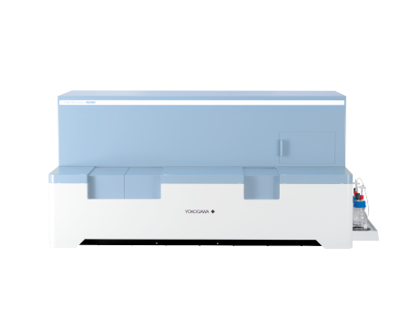
Single Cellome™ System SS2000
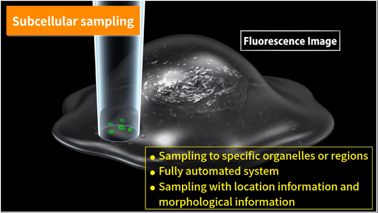
The SS2000 is a subcellular sampling system.
Automatically sample specific regions of cells or whole cells without any damages.
Image and analyze the cells with a confocal microscope simultaneously.
Detail
Brief Introduction
The SS2000 is a subcellular sampling system that automatically samples specific regions of cells or whole cells at the single-cell level while imaging cells in culture with a confocal microscope. The tip diameter is only 3-10µm, so avoid any damages to the cells. Because cells in culture do not need to be detached, positional and morphological information is maintained.


Product Schematic
Automated operation.
Precise pipetting control of location works.
Sampling with location and morphology information.
High resolution images and image analysis using confocal microscopy.
Incubator function for maintaining cell activity.

High usability samples
Samples can be collected on PCR plates and microplates and collect multiple samples in the same well. Samples can also be taken while being held in the glass tip without being ejected. The collection site has a cooling function to suppress sample degradation and an incubator function to maintain the culture environment. These samples can be used for genetic analysis, mass spectrometry, and single-cell cloning.

Work Procedure

1、 Intracellular components can be sampled at a single-cell level. This includes intracellular components that are difficult to sample by biochemical methods, such as organelles without lipid membranes.
2、 By sampling while maintaining the positional information, it is possible to sample and analyze the normal cells adjacent to the cancer cells and the normal cells located away from the cancer cells.
3、 Sampling is possible while maintaining morphological information. Allowing cells with different morphological changes can be sampled and compared.
4、 Different parts of a neuron, such as a cell body or axon, can be targeted for sampling.
5、 Single-cell cloning is possible from specific cells or cells with specific behavior under microscopic observation, such as transfected cells or virus-resistant cells. By combining various image analysis techniques, accurate and efficient cloning is possible.
6、 Multiple samples can be collected in the same well for analysis that requires a sample volume.
Application Fields
The SS2000 can be used for a variety of applications that require automatically sampling specific regions of cells or whole cells at the single-cell level.
Application
1、Subcellular sampling
Unlike existing cell isolation devices, the SS2000 can not only isolate a whole-cell but also sample only the target site inside the cell. It is possible to sample cytoplasm and regions containing target organelles selectively.
After staining of HeLa cell nuclei (blue), cytoplasm (green), and mitochondria (red), a mitochondria-rich region (arrow) of the cytoplasm was sampled.

2、Maintaining positional and morphological information of cells
Since only the target cells can be sampled without detaching the cells in culture, it is possible to sample while maintaining positional and morphological information.
Normal MDCK cells and green fluorescent-labeled abnormal MDCK cells were co-cultured at a 50:1 ratio. A normal cell adjacent to the abnormal cell exhibiting fluorescent signals (arrows) was sampled.

3、Live Cell Imaging with Confocal Microscopy
The SS2000 utilizes live-cell imaging products developed by Yokogawa. High-speed, high-resolution 3D imaging is possible using our unique confocal microscope technology. Samples can be taken from targeted cells under a confocal microscope in an incubator environment. Time-lapse photography is also possible, allowing dynamic changes in the target cell to be captured. Since it is possible to record moving images during sampling and images before and after sampling, it is possible to compare the results of analysis of collected samples with cell imaging data.
Target cells and sampling positions can be automatically selected by image analysis. (Targets can be automatically selected as shape of cells, size of nuclei, density of organelles etc.)

Video