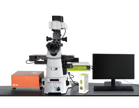
3D Organoid and Cellular Microenvironment Preparation and Fast Detection System
Innovative micron-scale customized organoid preparation tools
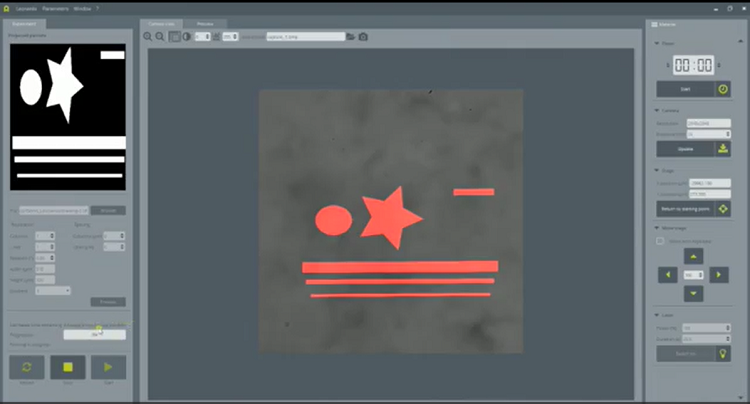
Organoids were prepared by contactless and maskless photopatterning method
With fast(25fps)and deep confocal imaging and detection
Suitable for organoids, organoid chips and other applications
Detail
Brief Introduction
In vivo, the cellular microenvironment has a crucial impact on the regulation of cell behavior and functions such as cellular differentiation, proliferation and migration. However, its biochemical and structural complexity limits our understanding of the key parameters regulating cell behavior.
In vitro, one of the challenges confronting cell biologists is therefore to work with controlled and reproducible microenvironments to more efficiently study living cells and model diseases.
Studying the influence of the microenvironment on intracellular and intercellular mechanisms has been essential for research in cell biology, for many years now. But in this quest, in vitro cell experiments confront researchers with many challenges, such as:
• the recurring reproducibility issues,
• reliability in term of physiological relevance,
• but also ease of use and efficiency.
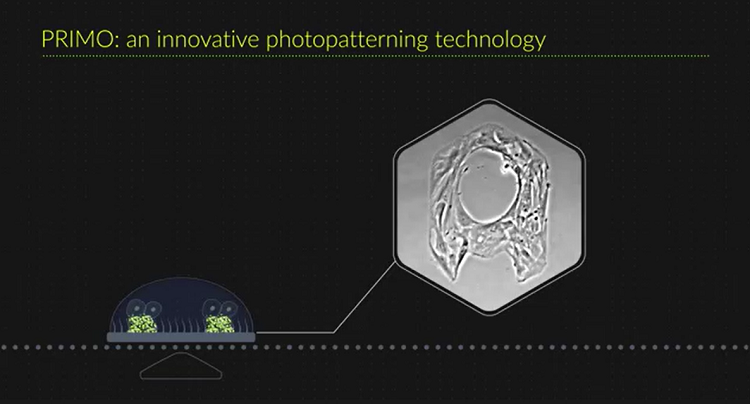
This is why our research team has developed PRIMO: a photopatterning system performing micropatterning, microfabrication and hydrogel polymerization to overcome all this issues. We make it evolves to a new generation, PRIMO 2, with compact footprint, optic schematics allowing fluorescence and faster performances.
After preparation of the organoid and other cell models, the system can also detect them with high speed (25fps)3D confocal imaging, suitable for long-term observation.

Product Principle
The system can create and prepare the organoid, organoid chip and other cell models through Micropatterning, Structuration of Hydrogel, Microfabrication and other mixed methods, and observe and detect them with fast(25fps)and deep 3D confocal imaging.
Please refer to the Product Principle of PRIMO and NL5.
Product Characteristics
Time saving & independence--Rapidly optimize your experimental conditions yourself.
High Flexibility--Download any images you want to structure and/or functionalize your substrate.
Versatility--Use your regular cell culture substrates: flat or microstructured, stiff or soft.
Protein micropatterning: glass coverslips, glass slides, 96 well plates, EM grids, polystyrene, PDMS, polyacrylamide gel (transfer), hydrogels.
Hydrogel structuration: UV-curable hydrogels, PEG Acrylate, Polyacrylamide, Agar, Matrigel.
Microfabrication: UV-curable materials, UV-photoresists.
More than 10 proteins used daily by our users, including: Fibrinogen-488, Fibrinogen-647, Fibronectin, GFP, Neutravidin-488, Neutravidin-647, PLL-PEG-Biotin, Protein A-647, Streptavidin, as well as primary and secondary antibodies.
The photopatterning resolution is 1.2µm.
The system can achieve multi-gradients etching with 256 gray levels.
The fastest etching time for a full field pattern of 500×300µm with 20× objective is 0.5s.
Fast scanning speed (25fps) for 3D confocal imaging.
High contrast deep 3D imaging without pinhole cross-talk.
Better resolution for deep 3D imaging than spinning disk confocal microscope.
16x brighter than spinning disk confocal microscope.
High sensitivity and resolution (170 nm after deconvolution).
Suitable for long-term live cell observation.
Application Fields
Organoid Applications:
1. PRIMO can create a 3D matrix microstructure through Hydrogel Structuration method, coupled with various cell growth factors necessary for organoid growth and development, to induce the iPSC (induced pluripotent stem cells) or ESC (embryonic stem cell) into various organoids with three-dimensional structure for functional research.
2. PRIMO can culture primary tumor cells from patients into tumor balls that are closer to the form in vivo for anti-tumor pharmacological experiments.
3. PRIMO can also create the 3D microstructures of Organ-on-chip or Organoid Chips by Microfabrication method to carry out co-culture of multiple cells, and simulate multi-cell microenvironments in vitro.
All other fields that study the interaction of cells with the microenvironment. Such as:
1. 2D cell microenvironment construction, such as cell migration/haptotaxis, cell adhesion, cell mechanics, standardized cell culture, standardized cell observation by electron microscope, etc.
2. 3D cell microenvironment construction, research such as tumor spheroid culture, organoids, tissue engineering, etc.
3. Microfluidic chip production, research such as organoid chip, cell fluid mechanics research, small molecule diffusion research.
Imaging and detection: Observe the Organoid, Organoid Chip and other cell models with fast (25fps) and deep 3D confocal imaging, suitable for long-term observation.
Application
1、Standard cell spheroid culture with non-adhesive matrix
Left panel: Schematic representation of a hydrogel spheroid assembly template with 4-arm-PEG-acryl hydrogel (blue). Right: Resulting hydrogel with growing HEK spheroids. Hydrogel microwells are photopolymerized with PRIMO. The 2nd row images are the confocal images of the cell spheroids.

PRIMO: Cell spheroids are the aggregation of cells that can serve as a physiological model or a starting model for complex organoids. The non-adhesive hydrogel matrix is solidified to form microwells, and the cells are planted to form cell spheroids, which can form 3D tissue organoid models with multi-cell coculture and multi-protein regulation.
2、Cell culture with adhesive matrix
Left panel: schematic representation of 4-arm-PEG-hydrogels with controlled heights (blue) patterned with Poly-L-lysin (red) and fibronectin. Middle: COS-7 cells seeded on the gels and stained for actin. Right: patterned molecules (red), actin cytoskeleton (green) and nuclear envelope (blue).
The 1st row images stimulate blood capillary, and the 2nd row images stimulate intestinal villi.

PRIMO: Mix different hydrogels to form an adhesive matrix, through photo-polymerization and photo-scission, let UV-sensitive hydrogels form a specific shape, the surface is functionalized, and then the cells are planted on it to simulate different organoids.
3、Functionalization of hydrogels
Combine protein micropatterning and hydrogel polymerization to turn common hydrogels into topographically and chemically heterogeneous environments.
The PRIMO projection system enables precise alignment of cells seeding on top of previously shaped hydrogels. Our technology permits the creation of substrate topology and allows a meticulous positioning of cells on the desire structure.


PRIMO: Mix inert hydrogels with other hydrogels to form an adhesive matrix, or modify the inert hydrogels to form an adhesive matrix, and then through photo-polymerization and photo-scission, the UV-sensitive hydrogels can form a specific shape, the surface is functionalized, and then plant cells to create different cell models, which can form 3D tissue organoid model with hydrogels as a skeleton and co-cultured with multiple cells and regulated by multiple proteins.
4、Organ-on-chip production
With the PRIMO technology, prototype your whole organ-on-a-chip in a record time and with a high reproducibility.
Rapid prototyping of microchips thanks to maskless photolithography.
Precise alignment of patterns with already existing structures to create multi-layer microfabrication.
Organ-on-chip production:
The PRIMO on-demand UV projection system is a powerful tool for fast-prototyping of microchips. With its highly flexible capacity, it is possible to craft new molds on a daily basis and use them as templates to create PDMS structures into which liquids or hydrogels can be flowed.
The examples down below show some chips designs with central and adjacent channels, and final PDMS chips made with PRIMO to simulate the bone microenvironment. Human Umbilical Vein Endothelial cells (HUVEC), Human Foetal Osteoblastic cells (hFOB) and Hematopoietic stem cells (HSC) were resuspended independently in collagen/fibrin hydrogel and injected in the different channels via the inlets. We illustrate the biocompatility of the chips by growing 3 different cell types in the different compartments for several days.

PRIMO: Design an "organoid chip" to create a co-culture microenvironment that mimics multiple cell interactions in vitro.
Video
PRIMO Introduction Video