PRIMO Bioengineering Custom Microenvironments System
Time saving & independence--Rapidly optimize your experimental conditions yourself.
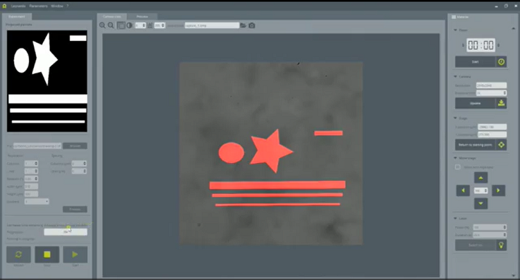
High Flexibility--Download any images you want to structure and/or functionalize your substrate.
Versatility--Use your regular cell culture substrates: flat or microstructured, stiff or soft.
Detail
Brief Introduction
In vivo, the cellular microenvironment has a crucial impact on the regulation of cell behavior and functions such as cellular differentiation, proliferation and migration. However, its biochemical and structural complexity limits our understanding of the key parameters regulating cell behavior.
In vitro, one of the challenges confronting cell biologists is therefore to work with controlled and reproducible microenvironments to more efficiently study living cells and model diseases.
Studying the influence of the microenvironment on intracellular and intercellular mechanisms has been essential for research in cell biology, for many years now. But in this quest, in vitro cell experiments confront researchers with many challenges, such as:
• the recurring reproducibility issues,
• reliability in term of physiological relevance,
• but also ease of use,
• and efficiency.
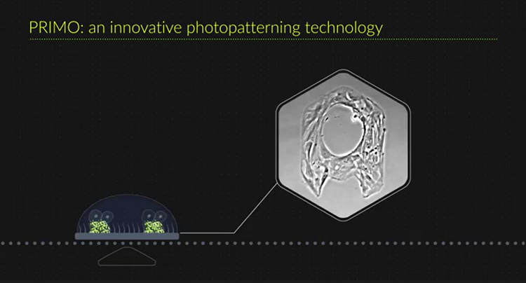
This is why our research team has developed PRIMO: a photopatterning system performing micropatterning, microfabrication and hydrogel polymerization to overcome all this issues. We make it evolves to a new generation, PRIMO 2, with compact footprint, optic schematics allowing fluorescence and faster performances.

Product Principles
The PRIMO bioengineering technology relies on a contactless and maskless photopatterning module (PRIMO) and a dedicated software (Leonardo). The platform can be used to structure cell culture substrates through photopolymerization and to functionalize any cell culture substrate surfaces (stiff or soft, flat or microstructured) through micropatterning.
1、Micropatterning:Surface functionalization
PRIMO micropatterning system allows to fine-tune cell adhesion to mimic in vivo phenotypes, study cellular mechanisms or isolate cells in reproducible conditions for standardized assays.
PRIMO photopatterning module allows to project on the substrate any image you want in order to create a protein micropattern. The combined action of UV (λ=365nm) and PLPP degrades the anti-fouling coating, in only a few seconds. The protein can then be added and will adsorb on the illuminated areas only.
Biomolecule Micropatterning method:

2、Substrate structuration
Use PRIMO photopatterning module to photopolymerize UV-sensitive resist or hydrogels and create structured substrates with controlled topography and stiffness. Micro-pillars, wells, grooves, PRIMO allows rapid prototyping of all the shapes you imagine to create substrates adapted to each experiment!
This method including:Hydrogel Structuration and Microfabrication.
Hydrogel Structuration method:


Microfabrication method:

3、Substrate structuration and functionalization
Whether on 3D substrates you manufactured yourself or on commercially available ones, PRIMO allows you to micropattern your biomolecules. Create more complex in vitro cell microenvironments and better 3D cell culture systems!
For an easier control over your experiments, Leonardo software automatically detects microstructures to precisely align and project your images onto it.
Complex Cell Microenvironments method:

Product Characteristics
Time saving & independence--Rapidly optimize your experimental conditions yourself.
High Flexibility--Download any images you want to structure and/or functionalize your substrate.
Versatility--Use your regular cell culture substrates: flat or microstructured, stiff or soft.
Protein micropatterning: glass coverslips, glass slides, 96 well plates, EM grids, polystyrene, PDMS, polyacrylamide gel (transfer), hydrogels.
Hydrogel structuration: UV-curable hydrogels, PEG Acrylate, Polyacrylamide, Agar, Matrigel.
Microfabrication: UV-curable materials, UV-photoresists.
More than 10 proteins used daily by our users, including: Fibrinogen-488, Fibrinogen-647, Fibronectin, GFP, Neutravidin-488, Neutravidin-647, PLL-PEG-Biotin, Protein A-647, Streptavidin, as well as primary and secondary antibodies.
The photopatterning resolution is 1.2µm.
The system can achieve multi-gradients etching with 256 gray levels.
The fastest etching time for a full field pattern of 500×300µm with 20× objective is 0.5s.
Application Fields
Modulate the biochemical and mechanical cues of the cell microenvironment in vitro with PRIMO and generate more physiologically-relevant cell models.
Primo system allows you to better study the behavior and development of living cells in a broad range of applications, such as: cytoskeleton dynamics, cell adhesion force measurement, cell confinement, cell migration, tissue engineering, spheroids.
Application
1、Cell force measurements
With PRIMO, it is possible to simplify measurements of cellular traction forces since it permits optical measurements of cell-mediated displacements to be converted into stresses and forces.
Down below, they performed cell force measurements in airway smooth muscle cells. They studied how extracellular matrix stiffness regulates force transmission pathways in multicellular ensembles of human airway smooth muscle cells.

2、Study Cardiomyocyte Contraction
With PRIMO, it is possible to pattern on any substrate’s stiffness, whether it is stiff or soft. It is also easy to combine micropatterning and hydrogel structuration to study cell forces.
In the example below, micropatterning was performed on soft and deformable substrates to study cardiomyocyte contractility. They observed that non micropatterned cells contract poorly and in an asynchronous manner whereas micropatterned cells show higher and more synchronized contractility.

3、Cell Migration
Rapid in-house optimization of custom micropatterns (adapted sizes, shapes, spacings)
Gradients or local control over protein density
Cell morphologies and migration velocities are regulated by the lateral confi nement. 
(g,h) Pattern composed of 5 interconnected microstripes of increasing widths: 5, 9, 13, 17 and 21 μm.
(i-k) The velocity increased from narrow to large microstripes (forth trajectory); then cells turned back
at the end of the larger microstripe and the migrating distance over time decreased from large to narrow microstripes (back trajectory).
4、Cell Adhesion Control
Substrates with modulated adhesion induce T cell haptotaxis. Cell tracking results on ICAM-1 (left panel) and VCAM-1
(right panel) substrates with modulated adhesion.
(A) Negative control on substrates with 0% adhesion contrast where cells migrated in random directions.
(B) Positive control substrates with 100% adhesion contrast where cells exhibited preferential haptotactic migration in the vertical quadrants (blue).
(C) Substrates with 50% adhesion contrast where cells haptotaxed in the vertical quadrants (blue) with an intermediate anisotropy index between
positive and negative controls. Each color represents the trajectory of migrating cell.

T cells migrating on patterned ICAM-1 substrates.

5、Cell positioning for Cryo-ET
Ease and improve whole Cryo-ET imaging experiments using the breakthrough PRIMO maskless micropatterning technology. Create perfect sample for FIB-milling and electron Cryo-tomography by directly controlling cell adhesion, localization and shape.
Higher yield of amenable cells for Cryo-ET experiments.
Precise cell positioning within EM grid meshes.
Multiple and standardized cell models.
Undamaged surface thanks to maskless photopatterning.

6、Tailoring 3D cell culture templates with common hydrogels
Left panel: Schematic representation of a hydrogel spheroid assembly template with 4-arm-PEG-acryl hydrogel (blue). Right: Resulting
hydrogel with growing HEK spheroids.

7、Cell culture of adhesive matrix
Left panel: schematic representation of 4-arm-PEG-hydrogels with controlled heights (blue) patterned with Poly-L-lysin (red) and fi bronectin. Middle: COS-7 cells seeded on the gels and stained for actin. Right: patterned molecules (red), actin cytoskeleton (green) and nuclear envelope (blue).

8、Functionalization of hydrogels
Combine protein micropatterning and hydrogel polymerization to turn common hydrogels into topographically and chemically heterogeneous environments.
The PRIMO projection system enables precise alignment of cells seeding on top of previously shaped hydrogels. Our technology permits the creation of substrate topology and allows a meticulous positioning of cells on the desire structure.


9、Organ-on-chip production
With the PRIMO technology, prototype your whole organ-on-a-chip in a record time and with a high reproducibility.
Rapid prototyping of microchips thanks to maskless photolithography.
Precise alignment of patterns with already existing structures to create multi-layer microfabrication.
Organ-on-chip production.
The PRIMO on-demand UV projection system is a powerful tool for fast-prototyping of microchips. With its highly flexible capacity, it is possible to craft new molds on a daily basis and use them as templates to create PDMS structures into which liquids or hydrogels can be flowed.
The examples down below show some chips designs with central and adjacent channels, and final PDMS chips made with PRIMO.

Video
PRIMO overall introduction video