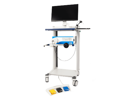
Fluorescence In Vivo Endomicroscope 2nd generation
The flflexible hand-held probe of the FIVE2
images can be captured at any angle with sub-micron resolution
Image resolution is 0.55µm lateral and 5.1µm Axial
vary image depth in the Z-Axis:up to 400 microns(tissue dependent)
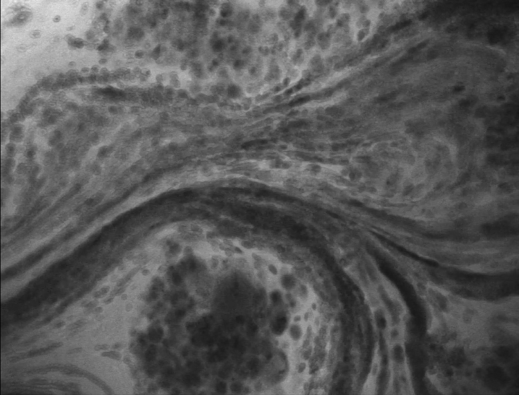
Thin optical sections enable reconstruction of 3D tissue structure in remarkable detail
Detail
Overall Introduction
The hand-held OptiScan FIVE2 system is the most advanced miniaturised confocal endomicroscope in the world.The FIVE2 enables you to push the boundaries of your research with maximum flexibility, viewing tissue at any angle with sub-micron resolution.

Product Principle
Optiscan technology is easy to use. Following application of a fluorescent dye, simply touch the probe against the tissue of interest and an image will be displayed on the screen in front of you.
A laser light passes through an optical fibre to a hand-held probe. Optics in the probe focus the laser to a diffraction limited spot in the tissue. Fluorescence is excited at this spot and the fluorescent light is captured into the optical fibre and passed to a detector. An image is generated by scanning the focussed spot through an image plane, then compiling the point intensity measurements. The image plane can be translated in depth, allowing generation of 3D image stacks.

Product Features
1.Slide-free and biopsy free:users can generate images at the sub-cellular level,in real-time.
2.Viewing live biological systems in-vivo at the cellular level in their entirety:not achievable through other imaging techniques.
3.Non-destructive:image creation is non-destructive to the tissue of interest.
4.Portable:easy to work on live tissues.
5.Image depth variation:unique ability to vary image depth in the Z-Axis:up to 400 microns(tissue dependent).
6.Zero cost image acquisition:just staining cost required.
7.Post-processing versatility:images can be easily post-processed to produce a range of presentations(incl 3D)
Why Choose ViewnVivo®?
Superb resolution:Using a distal end single scanning fibre rather than a proximally placed scanning fibre bundle provides unmatched lateral and axial resolution. The single fibre eliminates dead spaces and broken fibres, delivering high sensitivity and long lifespan.
Never miss an image:The ViewnVivo® imaging software includes an Image Rollback function. Users can select and save images from up to 60 memory held frames. Individual images or image selections can be saved or made into movie files.
Longitudinal studies:High level disinfection and sterilisation options provide a cost-effective, practical way to enable longitudinal studies on the same subject, as well as the ability to image cell cultures and other samples where sterility is required.
Long-life probes:ViewnVivo® probes are not a consumable item and will retain their imaging quality through years of operation and hundreds of procedures
Precise control in the Z-dimension:ViewnVivo® reports the position of the focal plane with micron accuracy. The imaging software provides an intuitive interface to set and collect Z-Stacks. This data can be turned into movies or ported into Fiji for processing
Image from any angle:The handheld flexible probe allows the user access to previously inaccessible samples.
Wide Range of Applications
Cancer research
•Tumour development/regression
•Metastasis
•Tumour detection/diagnosis
•Border demarcation
Tissue regeneration
•Stem cell research
•Orthopaedic tissue engineering
•Scaffold imaging
•Tissue rejection
Cell tracking
•Stem cell research
•Inflammatory responses
•Cell-cell & cell-tissue interactions
Photodynamic Therapy (PDT)
•Photosensitiser distribution
•PDT response
•Lesion detection & identification (PDD)
Infection & treatment
•Tissue microflora µbicide response
Pharmacology
•Drug delivery & pharmacokinetics
•Tissue response
Pathology
•Acute lung injury & animal models
•Liver disease models
•Morphological changes in disease
Microvascular research
•Dynamics and structure
•Angiogenesis/anti-angiogenesis
•Thrombosis
Animal models
•Tissue injury & disease
Molecular Imaging
•Receptor labelling
•Protein expression profiling
•Transgenic animals (eg GFP/YFP)
Application
1、Oral Cancer
Left: Normal
Middle: Pre-cancer
Right: Cancer
Optiscan technology captures sub cellular detail of oral mucosa in real time without having to do a biopsy. Scale bar =100um

2、Cervical Cancer
Left: Healthy squamous epithelium
Right: Cervical intraepithelial neoplasia
0.05%% acriflavine solution applied topically. Scale bar = 100um
Left: healthy colon
Right: Adenocarcinoma
Contrast agent: IV fluorescein sodium

3、Tumour angiogenesis
Left: Normal dermal microvasculature
Right: Melanoma-affected dermal microvasculature
In vivo CLE imaging shows changes in melanoma-associated vasculature in and around an implanted melanoma tumourof an athymic mouse. lntravenous administration of (0.3 ml, 10 mg per ml) FlTC-dextran was used as contrast agentscale bar = 100um

4、Autofluorescence
Left: orderly collagen fibres in ACL
Right: disordered collagen fibres after repeat loading of ACLAutofluorescence imaging of the anterior cruciate ligament (ACL). This study looks at ACL Failure from Fatigue RelatedMicrodamage. A collaborative project between Michigan University, Monash University and Optiscan Pty Ltd.Courtesy of Professor Mark Banaszak Holl - Engineering Department, Monash University
Scale bar = 100um

5、Lung endothelium triplel abelling
Contrast agent:
Lung endothelial cell nuclei: acridine orange, 0.05% i.v. (single arrow)Plasma: FITC-Dextran 70kDa, i.v. (double arrow)Distal epithelial membranes: FITC-R.Communis lectin (triple arrow)Scale bar = 100pm

6、Gastrointestinal microflora
Helicobacter in dog stomach.

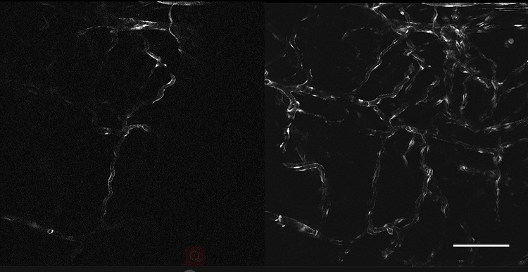
7、Zebrafish eye
Left: 180um Z-stack (Video) of Zebrafish eye expressing expressing endothelium specific gfp expression. Acriflavine wasused for background contrast.
Right: Maximum brightness projection of the Z-stack. Depth-colour coded in lmage ] Scale bar = 100um
左:成像深度:180μm,表达GFP的斑马鱼血管内皮成像,吖啶黄用作背景造影;
右:最明亮的一张,在Image J里深度颜色编码。Scale bar=100μm。
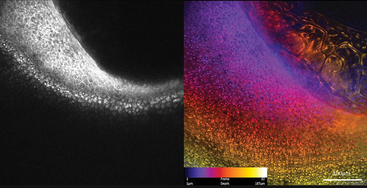
8、Brain cancer
Tumour island surrounded by leaky blood vessels in live rat brain. The tumour is a Glioblastoma which is highlyinfiltrative and causes hemorrhage. The contrast agent used here is intravenous fluorescein.Courtesy of researchers in Barrow Neurological institute, Phoenix, Arizona, USAScale bar = 100um
Video

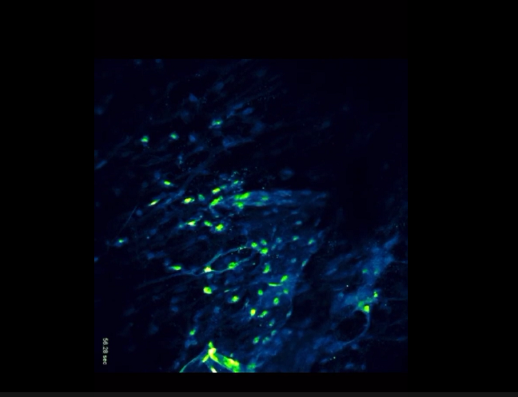
Tissue culture
GFP endothelium
Zebrafish eye
Brain cancer