
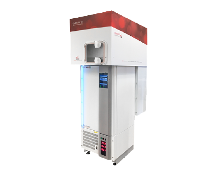
CellAssist 50 3D Organoid Culture and Quality Control System
Image adherent and suspension cells and 3D Organoid structures in 6- to 384-well flat bottom and round bottom plates
Automatically and consistently acquire and analyze 1,000’s of phase contrast and bright-field images at 4x, 10x, and 20x
Characterize cells and organoids with time series scans with 100+ focal planes 2 µm to 50 µm apart (user-selectable) with a z-range of 4.0 mm
High-throughput imaging with up to 50 microplates.
Detail
Brief Introduction
Thrive delivers instruments and software that provide previously unavailable automated live cell imaging and analytics for unparalleled insights into cell behavior, significantly advancing research and drug discovery.
CellAssist Family Takes Your Live Cell Imaging to 100+ Focal Planes.
CellAssist 50
Standalone, environmentally and robotically controlled 50-plate imager with each plate having a separately defined imaging schedule. Frequent, consistently timed, 24/7, remote imaging of up to 50 plates.

Product Characteristics
CellAssist Imager -- High-quality phase-contrast and bright-field imaging at 4x, 10x, and 20x in 6-well through 386-well plates.
Characterize cells and organoids with time series scans with 100+ focal planes 2 µm to 50 µm apart (user-selectable) with a z-range of 4.0 mm.
High-throughput imaging with up to 50 microplates (CellAssist 50).
CellAssist Documentation System -- Easily captures, with time-stamped barcode scans, critical information about researchers’ experiments.
Environmental Controls -- Temperature and gas controlled, (humidity with the CellAssist 50).
CellAssist Software and Analysis Workstation -- A suite of project set-up tools, secure datahandling, centralized database of projects and scan activity, and charting and analysis tools.

Application Fields
Growth & Characterization of Organoids & Spheroids
Studying & Screening Drug Response
Morphological Assays
Growth & Characterization of Stem Cells
Growth & Characterization of Suspension Cells
Tracking Single Cells
Counting Cells for Immunology in Co-Culture
Application
1、Organoid Study
Organoid Imaging with 100+ Focal Planes
Track organoid growth, morphological changes, and differentiation with excellent registration captured at multiple focal planes

Tracking a Stem Cell –based Differentiating Organoid
The Thrive instrument was superior to alternatives at a major research institution in order to track organoid growth, morphological changes, and differentiation with excellent registration at multiple focal planes.

2、Drug Screening Study
Measure Confluence Over Time to Measure Cell Health & Plate Consistency
Optimized confluence measurements for both colonies and non-colonizing cells

3、Wound Healing Research
Capture high-resolution, time-series images to screen dozens drugs quantitatively for measure wound healing.

4、Counting Two Cell Types in Co-Culture over Time
A major research institution for the first time could capture high-resolution, time-series images and use Thrive’s image processing capabilities to count two cell types based on differing morphology.

5、Tracking a Single Cell into a Colony
Capture high-resolution, time-series images to track single cell growth for label-free characterization of cell behavior.

6、Track and Characterize Spheroids
Monitor and track spheroid count, area, and morphology, for bodies up to 3.5 mm in size more accurately than other instruments.
iPSC Spheroids

7、Acquire Sharp and Complete Images of Suspension Cells
Acquire sharp images of all suspension cells in wells with fast imaging and up to 100 focal planes. Produced for a researcher for the first time sharp images of all the cells in all the wells.

8、Viral Plaque Detection without Staining
For the first time, researchers can characterize the formation of viral plaques and precisely follow their progression over time, and without using staining.
