
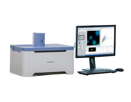
CellVoyager CQ1 Benchtop High-Content Analysis System
Easy to use, intuitive, simple acquisition software increases productivity
Compact design contains multiple, fully integrated functions
CSU W-1 confocal spinning disk technology results in higher scanning speeds and higher quality images
Systems open platform allows for integration for laboratory automation
Supports the CellPathfinder high content analysis system
Detail
Product Principles
Principles of the Microlens-enhanced Nipkow Disk Scanning Technology
A Nipkow spinning disk containing about 20,000 pinholes and a subsidiary spinning disk containing the same number of microlenses to focus excitation laser light into each corresponding pinhole are mechanically fixed on a motor, and very rapidly rotated. As a result, a high-speed raster scan of the excitation lights on the specimen can be achieved. The pinhole and microlenses are arranged on each disk in our proprietary design to optimize the raster scan. Multi-beam scanning not only increases scanning speed but also results in significantly lower photobleaching and phototoxicity because multi-beam excitation needs only a low level of laser power on the specimen to fully excite fluorescence.

Brief Introduction
With its compact footprint and lightweight, benchtop design, there is no need for a darkroom or specialized setup for the CellVoyager CQ1. The unit provides rich feature extractions, facilitating sophisticated cellular image analysis, and while the Nipkow Spinning Disk Confocal Technology allows high speed scanning, it reduces phototoxicity and photobleaching.

Product Characteristics
Enables measurement of spheroids, colonies, and tissue sections
No need to remove cells from the culture dish, in contrast to traditional flow cytometry
Nipkow spinning disk confocal technology allows high-speed yet gentle 3D image acquisition
Rich feature extraction to facilitate sophisticated cellular image analysis
Wide field of view and tiling capability enables easy imaging of large specimen
Enables analysis of time-lapse and live-cell
High precision stage incubator and low phototoxicity of our confocal makes the analysis of time-lapse and live-cell are possible.
Max. 20fps option for fast time-lapse.
High-quality image and similar operability to a traditional flow cytometer
Integration with CellPathfiner software can provide powerful analysis displayed in real-time with image acquisition.
Usable high-quality image as confocal microscope image.
Interactive graphs make it easy to trace back the data points.
Open platform
Connectable with external systems via handling robot.
Expandable to the integrated system as image acquisition and quantification instrument.
FCS/CSV/ICE data format readable by third-party data analysis software.
A variety of cell culture and sample dishes are applicable.
Compact footprint, lightweight bench-top device; no need for a darkroom.
Application Fields
Typical application areas include:
Cell biology research;
Organoid research;
Neurobiological research;
Stem cell research
Drug screening
Application
1、3D Measurement
The CQ1 is the easiest way 3D measurement system. Simple cell identification, colony counting, and complex colony property analysis are available. Of course you can do whole well imaging and analysis。
Example 1:Quality Control: Aggregated cell images were taken in slices and presented as 3D. Marker expression level as well as spatial information of individual cells were quantified via image analysis.
Template: Spheroid structure: Cell-by-cell measurement of aggregated, cells like spheroids.
Applications: Sheroids, Differentiation

Example 2:Quality Control:Sphere shape and pluripotencymarker expression level are suitable index for evaluation of quality of pluripotent state of human iPSC sphere.
Template: Colony measurement: Cell-by-cell measurement of aggregated, cells like spheroids.
Applications: Colony growth evaluation, Differentiation.

2、High Content Analysis
High quality confocal images from the CQ1 can be used for more detailed image analysis. Morphology change, particle analysis and other. High Content Analysis that require high resolution images. Of course the CQ1 can work like simple Confocal Microscopy to get a nalyzed data and images.
Example 1: Analysis: gamma-H2AX focus formation
The phosphorylation of histone H2AX Ser139 (gamma-H2AX) is one of the significant events upon the breakage of double strand DNA. Quantitative measurement of gamma-H2AX focus formation can be easily performed by using the high-speed confocal image acquisition in combination with the Granule Analysis Template.
Template: Dots in Nucleus: Measurements of dots in cytoplasm and nuclei, Precise separation of individual dots by the confocal unit.
Applications: FISH, GPCR.

Example 2: Nuclear translocation
NFκB is one of the famous transcription factor of DNA. NFκB plays a key role in regulating the immune response and inflammation and is attracting attention as a tumor therapy and anti-inflammatory drug target. NFκB is located in the cytoplasm with IκB which is inhibitory protein. Once the signaling pathway has been activated by the cytokine stimulation via cell membrane receptor, dissociate IκB from NFκB and activate NFκB. Then NFκB translocate into the nucleus to bind specific sequence of DNA, which induce inflammation. Nucleus and intracellular NFκB level indicates protein level between cytoplasm and nucleus.
Template: Nucleus and Cytoplasm: Measurements of nuclei and cytoplasm, Precise separation of localization by the confocal unit.
Applications: Nuclear translocation, Membrane translocation

3、Time Lapse Imaging
Keep cells happier in incubator to see how they react on live. The CQ1 is installed with Yokogawa's proprietary technology CSU, which is very gentle to cell-friendly, low photobleaching and low phototoxicity. Long-term time lapse are possible while minimizing the effects of multiple measurements.
Example1:Time lapse analysis: Apoptosis
Spread HeLa cells to 96well microplate with 10,000 cells/well. Stain with Hoechst 33342 (1 µg/ml, 30 min, 37 °C) and treat with Staurosporine (0 - 10µM) and capture image every 15 min. Recognize DNA fragmentation area of nuclei at Staurosporine 10 µM treatment.
Template: Nucleus: Measurements of Volume, Intensity and Morphology
Applications: Cellcycle, Apoptosis.

Example 2: Time lapse analysis: ESC colony
Time lapse analysis of colony size and individual cells allow to monitor colony formation state. CQ1ʼs image can perform image acquisition with low photo-toxicity.
Template: Colony measurement: Time course measurement allows to monitor cell colony growth.
Applications: Cell colony growth, Differentiation.

4、CTC (Circulating tumor cells)
CTCs are tumor cells which circulate in peripheral blood. Developed tumors metastasize through the bloodstream and lymph fluid. Therefore, tumor cells exist in the bloodstream when metastasis occurs. The detection of CTCs makes it possible to diagnose recurrence and metastasis at an early tumor stage. CTCsʼ numbers are very small as only less than 100 CTCs are contained in more than 1x10⁶ of blood cells in 10 ml of cancer patientʼs blood. Therefore it is difficult to detect CTCs with a flow cytometer because they detect CTCs as noise. However, it is very easy to detect rare CTCs with an Image cytometer.
Template: CTC: You can detect multiple marker expression of the cell. Not only for circulating tumor cells, but also for the other specific marker can be detected.

5、Cell cycle analysis: M-phase inhibitor
Cell cycle analysis in relation to H3Ser10P immunofluorescence by utilizing the CQ1ʼs multi-color channel capabilities. Histone molecules are phosphorylated during cell cycle progression with phosphorylation of the 10th serine of histone H3 being one of the well characterized events of late-G2 to M progression.
Template: CellCycle: You can detect cell cycle to verify drug treatment efficiency. This is available by the flow cytometer, but CQ1 can analyze more items which typical at the image cytometer.

6、CellPathfinder software
The dedicated analysis software "CellPathfinder" can analyze a large amount of image data from multiple angles and easily lead to a graphical display. In addition to the machine learning function, the new Deep Learning option dramatically improves the recognition of analysis targets, making it powerful not only for bright field image analysis, but also for difficult analysis such as 3D culture systems and live cell imaging.
