
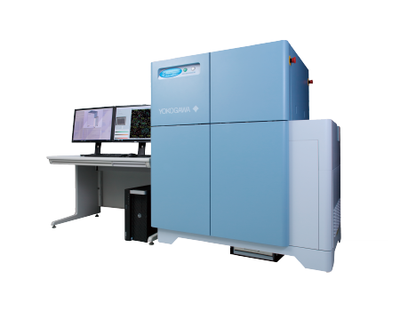
CellVoyager CV8000 High-Content Screening System
Spinning Disk Technology result in higher scanning speeds and higher quality images
Real time Confocal, label-free imaging for Live and Kinetic experiments
High throughput increases efficiency
Reliable, proven technology
Detail
Product Principles
Principles of the Microlens-enhanced Nipkow Disk Scanning Technology
A Nipkow spinning disk containing about 20,000 pinholes and a subsidiary spinning disk containing the same number of microlenses to focus excitation laser light into each corresponding pinhole are mechanically fixed on a motor, and very rapidly rotated. As a result, a high-speed raster scan of the excitation lights on the specimen can be achieved. The pinhole and microlenses are arranged on each disk in our proprietary design to optimize the raster scan. Multi-beam scanning not only increases scanning speed but also results in significantly lower photobleaching and phototoxicity because multi-beam excitation needs only a low level of laser power on the specimen to fully excite fluorescence.

Brief Introduction
Within the drug development market, demands on High Content Analysis systems for drug efficacy evaluation are increasing in accordance with the needs for cell-based assay and phenotypic screening. In order to increase screening efficiency, devices with higher speeds (higher throughput) are required.
On the other hand, in order to bridge the “valley of death” of the drug development process, the quality of screening hits must be increased.
This requires the construction of more complex evaluation systems that utilize multifaceted parameters via 3D cultivation systems, live-cell imaging and higher detail image analysis.
In current drug development research, determining how to implement throughput screening and complex evaluation system screening in parallel is an important issue.
The CellVoyager CV8000 is a high-end, High Content Analysis system that solves this contradictory screening challenge. Through the combination of a proprietary Yokogawa High Speed Confocal Scanner, water immersion lens, up to four high field-of-vision cameras, a microscopic stage with cell cultivation environment, and an integrated robotic pipetter, we have realized not only high throughput, high-resolution imaging, but also phenotypic screening via a more complex evaluation system. In addition, our specialized analysis software, CellPathfinder, uses deep leaning and machine learning to recognize target objects with high accuracy, supporting you from image analysis to results display using graphs.

Product Characteristics
1、Dual spinning disk confocal system
A Yokogawa proprietary multi-scan method utilizing approximately 1,000 laser beams on the observation region and tandem disks rotating at high speed. The disks comprise a pinhole array disk with approximately 20,000 pinholes arranged in an equal pitch spiral pattern, and a microlens array disk that focuses the excitation light laser into individual pinholes. Not only does this allow high speed imaging, but it also largely prevents phototoxicity and fluorescence photobleaching. Please refer to the Product Principle.
2、Pinhole disk exchanger: deeper,clearer observation
Two different types of pinhole disks (25/50μm) can be used, according to the sample. For thick samples, reducing the pinhole diameter allows for higher confocality, shaper images. For dark samples, increasing the pinhole diameter allows for brighter images.

3、Optical configuration: higher throughput screening
The optical system configuration can be selected according to the purpose. A single 96-well plate can be imaged in four colors in one minute by attaching four high- sensitivity wide-field sCMOS cameras. The system is also compatible with FRET and CellPainting assay.

4、Original water immersion lens: Capturing finer structures
Water immersion lenses excel in capturing high-resolution images of cells within a liquid. The CV8000 can be equipped with a 40x or 60x water immersion objective lens. Our 40x lens is a particularly unique lens capable of highly advanced spherical aberration correction, allowing for the capture of bright high-resolution images over a full wide-field. The lens water supply is also completely automated. This equipment makes high throughput screening via water submersion lens possible.

5、High-precision incubator and robot pipetter: Capturing live cell movement
The stage incubator features an airtight construction, managing humidity, temperature and CO2 levels. The integrated robotic pipetter conducts the following process fully automatically: tip pickup → reagent collection from the reagent plate → reagent addition to the sample plate →tip disposal. Not only can images be rapidly obtained before and after reagent instillation, but it’s also possible to add reagents to single wells multiple times, and adjust the addition speed etc., broadening the range of dynamics observation via reagent instillation.

6、Featuring a s table built-in stage incubator: Making long-duration live-cell imaging possible
HeLa cells were seeded in a 96 well plate at a density of 500 cells per well, and cultured for 24 hours. The well plate was then placed in the internal stage incubator and cell culturing was conducted for 72 hours, and the total area (hereinafter Total Area) occupied by cells was analyzed. As a result, minimal unevenness in cell multiplication was observed across the 96-wells (excluding the four corner wells) when compared to a regular CO2 incubator.

7、System Integration
Centralized process management, from the cultivation environment, to transfer, imaging, analysis and data management. We offer optimum systems in response to our customers’ needs.

8、High Content Analysis Software: CellPathfinder
The software analyzes image data captured with the CV8000, creates graphs and exports various data. Beginner and expert users alike can take full advantage of the software, thanks to an abundance of templates and flexible protocol editing capability. CE bright field and machine-learning functionalities make label-free analysis possible. The new Deep Learning option has also been added, largely improving cell recognition accuracy.

Application Fields
Applications include drug screening and optimization, pathway analysis, toxicity testing, cell-based large-scale screening, and cell quality control etc.
Application
1、Long-term Live Cell Imaging
Stage incubator included as standard. Realization of non-stop, long-duration observation (3 days +) via humidity, temperature and CO2 control.

2、Kinetic Assay
Drug addition during imaging is made possible by an integrated robotic pipetter with disposable tips. Ideal for kinetic experiments involving the observation of high speed phenomena.

Ionomycin concentration-dependant calcium response
It’s possible to conduct high speed imaging before and after ionomycin instillation, recognize individual cells from the images, and obtain timelapse data for each cell.


3、Organoid/Spheroid
Yokogawa′s spinning disk confocal technology excels in imaging of samples with depth, such as 3D culture samples where clear and quick imaging is difficult, allowing for evaluation close to in-vivo quality.

4、Label-free analysis
Recognition and analysis can be performed by taking bright field images from several Z positions and creating a CE bright field image using the included CellPathfinder analysis software. Analysis accuracy is further enhanced via the new Deep Learning option.
