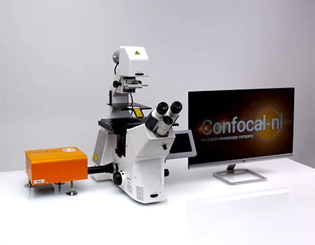
NL5 Fast & Deep 3D Confocal Microscope
Using slit apertures to construct a confocal imaging system
Fast scanning speed with low phototoxicity and photobleaching
Better resolution for deep 3D imaging & 16x brighter than spinning disk
Suitable for long time imaging of living samples
Excellent add-on to any widefield microscope.
Detail
Product Principle
Spinning disk systems are great but when going deeper they all show pinhole crossover issues causing image quality degradation. Also they need “help” to create the uniformity of illumination.
NL5 is using slit apertures, rather than pinholes, to construct a confocal imaging system, which has some advantages. The signal level is increased and, if a detector array is used, a line image can be generated in real time. Slit apertures can also be used in a direct-view confocal (tandem scanning) microscope.
Convert your existing research microscope into fast scanning confocal system ideal for deep 3D live cell imaging with NL5.
Brief Introduction
NL5 is a fast line-scanning confocal system with high sensitivity and resolution. It allows you to quickly screen a multi-well plate with multicolor images, from which you can select the most promising ones. Capture 3D live samples with minimal phototoxicity, high resolution and contrast. Like RCM, NL5 keeps your samples happy under the microscope for a long time and is easy to use. Convert your research microscope into a fast-scanning confocal microscope that provides very gentle conditions for your live samples.

The Benefits of NL5
Single Digital scanner (same type as RCM2)
Faster scanning speed than RCM2
High contrast deep 3D imaging without pinhole cross-talk
Better resolution for deep 3D imaging than spinning disk
16x brighter than spinning disk
High sensitivity and resolution (170 nm after deconvolution)
Hardware integration into third-party software
Extreme sensitivity ensures low phototoxicity, live cell friendly
Compact & easy to use
Fast scanning speed, low levels of phototoxicity:
In the field of biology, some natural processes happen fast, like cell and protein dynamics and cell-cell interactions. Therefore, fast scanning speed is needed to study them. And that is exactly what NL5 brings to the table! In addition, researchers in developmental biology, regeneration or immunology fields often need to image large live samples (mouse embryos, zebrafish, organoids) for a long time without losing any detail of the cell’s behavior. In that case, a combination of speed and a low phototoxicity system like NL5 is perfect.
The benefits of using NL5:
NL5 is a fast scanning confocal system that reaches 55 fps (full frame), much faster than regular point-scanning confocal systems. In addition to speed, it brings very gentle imaging conditions for your samples as well. Perform live cell imaging for more than a day, with no signs of phototoxicity. NL5 is easy to use, and requires basic training to operate.
NL5 does not cause any pinhole cross-talk, as it has only one slit pinhole. Pinhole cross-talk is associated with spinning disk systems, because they contain a disk with multiple pinholes combined and the out-of-focus light can sometimes pass through the closeby pinholes. This effect is more evident when imaging deep within the sample, making NL5 the ideal system for those applications.
NL5 is a cost-effective solution that can be coupled to any research microscope via the c-mount interface.
Applications for NL5
NL5 is an easy-to-use and compact confocal microscope system, a great addition to any microscopy facility. It’s a fruitful solution for individual research labs to easily convert any existing widefield fluorescence microscope into a fast confocal microscope system with high sensitivity and resolution. Capture datasets of 170nm lateral resolution after deconvolution at 55 fps. Study fast cell and molecular dynamics, or perform high contrast deep 3D imaging with minimal aberrations. The high sensitivity of the modern sCMOS cameras used as detectors facilitates acquisition with low light sample exposure, making NL5 ideal for live cell imaging.
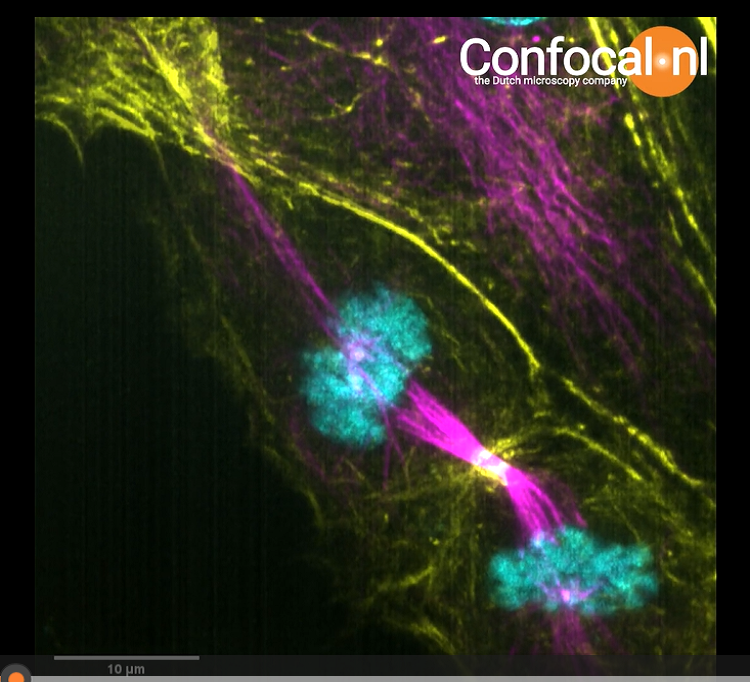
1、Fast cell dynamics
2、Live cell imaging
3、Deep 3D imaging
4、NL5 imaging with deconvolution
5、High content screening
Application
1、Fast cell dynamics
Study fast cell dynamics with very low phototoxicity, such as Cell Migration, Intra / Extra-cellular Transport, Cell Signaling. Better visualization and analysis of thin and dynamic structures even with dim signals.
HeLa Cell with cytosolic GFP (video):
2、Intestinal tumor organoids in mice
Long-term NL5 imaging for intestinal tumor organoids in mice (video):
3、Live cell imaging
Long-term live cell imaging in gentle conditions, low phototoxicity and bleaching, such as Developmental Biology, Regeneration, Immunology. Imaging happy cells and organisms for more than a day.
Zebrafish embryo (24 hpf). Endothelial cells labelled with GFP
z-stack (500 slices) every 10 min for 24 hours (video)

4、Deep 3D imaging
Deep 3D imaging, such as Organoids, Zebrafish / mouse embryos, 3D culture with hydrogels.
NL5 vs NL5 (deconvolution) vs SDC (Spinning disk confocal):
Spinning disk: pinhole cross - talk aberrations in deep frames
NL5: 1 slit pinhole - better images in deeper frames
A Nikon TiE microscope was used with CFI Plan Apo Lambda 100X 1.45NA oil objective. Sample: Volvox, autofluorescence was excited using a 561 nm laser.

The NL5 images are much brighter and have better resolution than Spinning disk confocal images at 60µm deep and 90µm deep:


NL5 image at 90μm deep:

5、NL5 imaging with deconvolution
NL5 original image vs NL5 deconvolution image:
The image after deconvolution is sharper and has higher resolution.

Video

Fast cell dynamics-HeLa Cell with cytosolic GFP
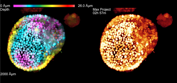
Long-term imaging of intestinal tumor organoids in mice
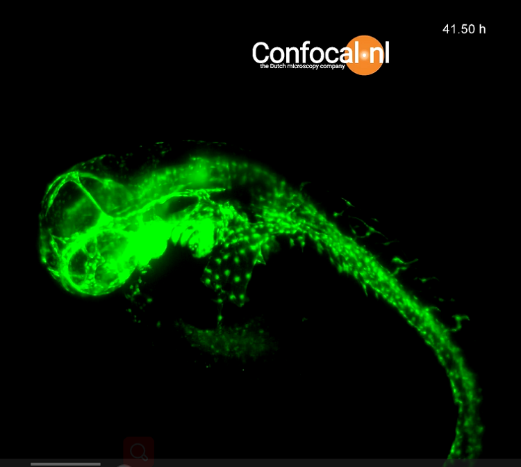
Long-term live cell imaging-Zebrafish embryo