
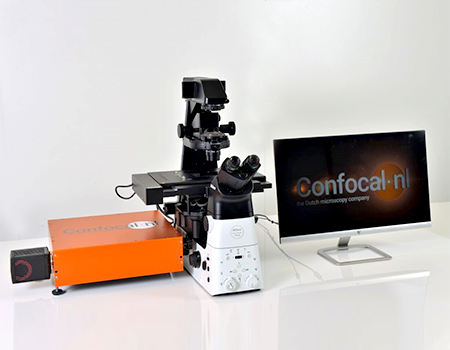
RCM Re-scanning Confocal Microscope
Super-resolution (120nm) imaging with 40x 1.4NA objectives.
Higher sensitivity and higher signal-to-noise ratio
Low phototoxicity and photobleaching, suitable for long time imaging of living samples
Larger FOV without increasing acquisition time
Excellent add-on to any widefield microscope.
Detail
Product Principle
RCM adds the combination of Unit 1 (Confocal microscope) and Unit 2 (Re-scanner + CCD) to a conventional microscope and updates it to be a higher-resolution confocal microscope. It can capture datasets of 170nm resolution raw (120 after deconvolution) over a very large FOV. Study fast live-cell dynamics and perform 3D imaging in optimized conditions.
Principle illustration:

Compatible microscope:

Brief Introduction
Sharp images, a large field of view and easy to use? It’s possible with Confocal.nl. Get more from your samples, with a higher resolution and a higher sensitivity than conventional confocal microscopes and study live samples longer. Our Re-scanning Confocal Microscope (RCM) uses multiple laser pointers and a camera as a detector. The low laser power required is live cell friendly: it prevents phototoxicity in your live samples and photobleaching of your fluorophores. Creating long-term time lapses at a super-resolution was never this accessible.
RCM2 is our second-generation RCM, with digital scanner technology. It makes re-scanning the standard and allows a speed of 2fps at 512×512 pixels. RCM2 has optics to make it suitable for super-resolution imaging with high NA objectives in the low magnification range, like 40x 1.4. A lower magnification allows for a bigger field of view (FOV), brighter images, and even lower laser power. RCM2 has demonstrated imaging at 10 nano-watt excitation power!
Improvements of sCMOS cameras allow you to sample resolution of low magnification objectives effectively, without increasing the exposure time. In a regular PMT-based confocal this is not possible.
The Benefits of RCM2
1、Using RCM2 with a 40x 1.4 objective, you can see more cells at full resolution at once. A larger field of view increases the chances of getting the results you need.
2、Obtain sharp images with a high signal-to-noise ratio even in samples with a low amount of epitopes or weak stainings. Get more from your samples.
3、Use even lower laser power to minimize phototoxicity and photobleaching during live-cell imaging.
4、Getting super-resolution raw images, without averaging or integration, reduces the acquisition time and allows for a more precise analysis of the subcellular structures.
Wide Field image vs RCM image:

Applications for RCM2
Capture datasets of 170nm resolution raw (120nm after deconvolution) over a very large FOV. Study fast live-cell dynamics and perform 3D imaging in optimized conditions. The increased detection efficiency facilitates acquisition in low fluorescence conditions, like single-molecule detection (smFISH). In combination with silicon objectives, RCM2 allows for deep 3D imaging of organoids, zebrafish embryos, or larger live samples.
Application
1、Getting Super-resolution Raw Images
Deer skin fibroblast cells as below, stained for: DAPI (blue), Phalloidin-Alexa488 (green), Mitotracker CMXRos (red).
RCM can obtain 170nm resolution raw and 120nm after deconvolution.

2、2D Cell Imaging
2.1 Neurons in co-culture stained for Actin (red), MAP2 (magenta) and Tau (green). Imaged on RCM2 with a 40x 1.4 objective.

2.2 Drosophila Melanogaster heart tube stained for Actin (green), Myosin Heavy Chain (red), Alpha-tubulin (blue). Imaged on RCM2 using 40x 1.4 objective.

2.3 HUVECs cells stained for Tubulin (yellow), Actin (purple) and DNA (blue), Imaged on RCM2.

2.4 BPAE cells stained for Actin (green), Mitochondria (red), DNA (blue). Imaged on RCM2 using 1 40x 1.4 objective.

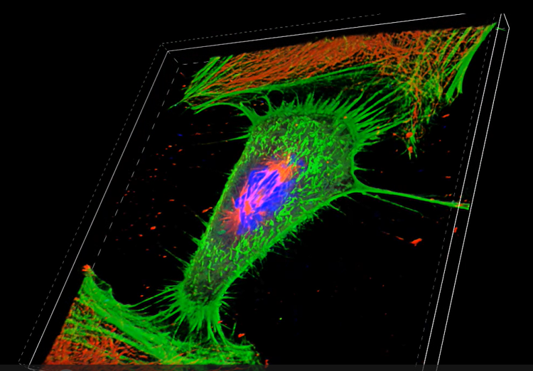
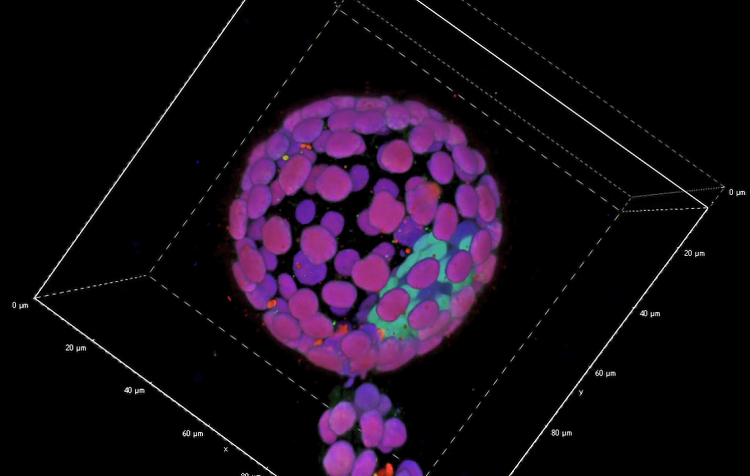
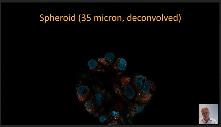
3、3D Image Reconstruction
3.1 Hela Cell in Mitosis, stained for Actin, Cytoskeleton (green), Tubulin, Microtubule (red), DAPI, Nucleus (blue).
3.2 Mouse Blastocyst, stained for GATA4, primitive endoderm marker (green), CDX2, trophectoderm marker (red), DRAQ5, Nucleus (blue).
3.3 Tumor Spheroid, stained for DAPI, Nucleus (blue), Spheroid, 35um, deconvolved
4、Long-time Live Cell Imaging Mitochondria Dynamics
Cell type: HOIN1 cells
Laser power: 1 µW
Framerate: 1 frame per 10 seconds
Duration: 61hr06m
Total: 22,000 frames
After 61hr06m, it was observed that the cells were still alive and still dividing, indicating that the laser had little toxicity and little damage to living cells.
Video
3D Hela Cell in Mitosis
3D Mouse Blastocyst
3D Tumor Spheroid
