
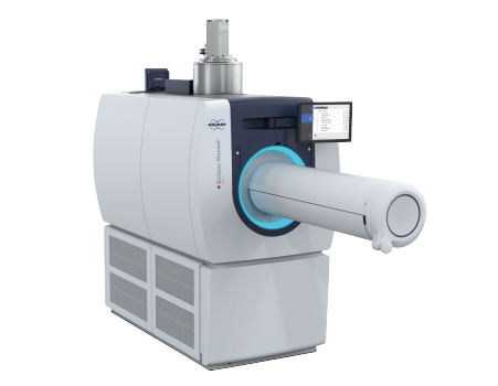
Bruker BioSpec Maxwell Intelligent Preclinical MRI
Maxwell Magnet Technology
No liquid helium filling combined with 3 Tesla, 7 Tesla, and 9.4 Tesla field strengths
Self-monitoring
Push button auto-cooling and auto-charging for maximum uptime
Compact
Small footprint, self-shielded magnet for maximal siting possibilities in small and/or BSL areas
Detail
Maxwell Magnet Technology
Maxwell magnet technology makes superconducting Magnetic Resonance Imaging without the need for liquid cryogen filling possible. This revolutionary magnet technology is based on recondensing cooling of a minuscule volume of helium and a patented cooling technology . A powerful dual stage cold head maintains the low temperature of < 4.2 K, allowing constant recondensing of the helium, which in turn cools the superconducting coil.
Features superconducting magnet conduction cooling technology
Eliminates liquid cryogen filling and the need for quench pipes
Simplifies siting and maintenance
Incorporates automatic self-supervision
Empowers push-button magnet control
Enables most demanding studies
Brief Introduction
BioSpec Maxwell MRIs recreate preclinical MRI, taking operational ease of use to a new level. Cutting-edge autonomous instrument control continually self-regulates instrument parameters to their optimal state, providing a completely new ownership and imaging experience.
The BioSpec Maxwell MRI’s innovative Maxwell magnet technology eliminates the need for liquid helium filling, providing long-term cost savings and making use inherently safe.
With their small footprint and minimal site requirements, the BioSpec Maxwell MRIs perfectly fit into existing lab structures, expanding biomedical research capabilities with easy-to-use MRI.
The intuitive workflows ensure highest productivity for a wide range of preclinical MRI studies on small rodents.
While this simplicity guarantees best results, the flexibility of classic BioSpec features such as the method development platform and the possibility to add an MRI CryoProbe or PET module provide users with the unparalleled versatility and full range of freedom that is needed to push research boundaries.
Product Characteristics
1. Ease of Use and Maximal Productivity
Accurate animal positioning with the motorized Animal Transport System, including touchscreen operation even with gloves for a simplified, precise workflow
Intuitive ParaVision software with over 100 validated and ready to use in vivo protocols and scan programs for mice and rats
Streamlined workflows including automatic quantification
Safe operation with inherent shielding
2. Maximum Cost Savings
No liquid helium or nitrogen fillings provides freedom from fluctuating helium prices
No Faraday cage required for easiest siting
No quench pipe required for easiest siting
Lightweight and compact footprint fits in any laboratory
3. Maximal Experimental Freedom
3T, 7T, and 9.4T
Complete RF coil portfolio for mice and rats available including support for very large rats with best-in-class free RF-coil access of 82 mm
ParaVision software with more than 1,000 sequence variations including IntraGate based methods, UTE, and ZTE
Method development platform
Most advanced pulse-tube cold head for minimal vibrations for sensitive studies such as fMRI
Gradient strength up to 900 mT/m, Slew rate: 4200 T/m/s
Upgradable with MRI CryoProbe delivering an exceptional sensitivity increase
Upgradable with state-of-the-art PET insert or PET inline module
4. Maximal Reliability and Up-time
Hold-time minimum of 6 hours during cooling disruption1
Extensive sensors monitoring of instrument parameters
One-click charging and discharging
One-click cooling and warming
Application
1、Oncology
Spectroscopic imaging of hyperpolarized pyruvate and lactateconversion reveals intertumoral variation in patient derivedrenal cell carcinoma. 2D EPSI at 3T after infusion ofhyperpolarized [1-13c] pyruvate in mouse model.

2、Drug Development
CEST imaging at 3T enables monitoring of compositionachanges of hydrogel-based drug treatment for glioblastomamultiforme (GBM), providing valuable insights for treatmentrefinement.

3、Neurological Studies
FLASH angiography, such as this rat brain study at 7T can beused to investigate arterial abnormalities such as stenosisocclusions and aneurysms. Resolution in plane: (94 x 94)um2, Slice Thickness: 400 um, Slices: 80, Acquisition time: 08m 15 s

Rat brain RARE at 7T, segmentation with pmod
